Categories

IT band syndrome affects runners and athletes, causing pain on the outside of the knee. Understanding the condition is key to effective recovery and prevention.
The iliotibial (IT) band is a thick band of fibrous tissue that runs along the outside of your thigh, extending from the hip to just below the knee. It’s not actually a muscle, but a strong, dense connective tissue called fascia.
Its primary function is to stabilize the knee and hip during movement, particularly during running and walking. It works with several muscles – including the gluteus maximus, tensor fasciae latae (TFL), and quadriceps – to provide support and control leg motion.
Think of it as a rope connecting these muscle groups. While naturally somewhat tight, the IT band’s flexibility can be influenced by the muscles it connects to. It plays a crucial role in preventing the knee from buckling inwards and assists in hip abduction and external rotation. Understanding its anatomy and function is vital when addressing issues like IT band syndrome, as treatment often focuses on the surrounding musculature.
IT Band Syndrome (ITBS) is a common condition causing pain on the outside of the knee, particularly among runners and cyclists. It’s not inflammation of the IT band itself, as previously thought, but rather friction between the IT band and the bony prominence of the femur (thigh bone) during knee bending and straightening.
This friction irritates a bursa – a fluid-filled sac – located under the IT band near the knee. Over time, this irritation leads to pain and discomfort. The pain typically worsens with activity, especially downhill running or cycling, and may subside with rest.
ITBS isn’t usually caused by a sudden injury, but develops gradually due to repetitive stress. Factors like improper training, inadequate stretching, muscle imbalances, and biomechanical issues contribute to its onset. Early diagnosis and appropriate management are crucial to prevent the condition from becoming chronic.
Several factors contribute to ITBS, including anatomical variations, biomechanical inefficiencies, training intensity, and inadequate warm-up routines before physical activity.

Leg length discrepancies, even subtle ones, can significantly impact IT band tension. A shorter leg on one side often leads to increased stress on the IT band of the longer limb, as the body compensates during movement. Similarly, external tibial torsion – an outward twisting of the shinbone – can alter biomechanics and contribute to ITBS. This rotation can increase friction as the IT band rubs against the lateral femoral epicondyle.
Varus alignment of the knee, where the knees angle inward, is another predisposing anatomical factor. This alignment increases the load on the outer aspect of the knee, exacerbating IT band stress. Foot structure also plays a role; individuals with high arches or excessive pronation may experience altered lower limb mechanics, contributing to ITBS development. Finally, the shape of the femoral epicondyle itself can influence friction – a more prominent epicondyle increases the likelihood of IT band irritation. These anatomical predispositions don’t guarantee ITBS, but they heighten susceptibility when combined with other risk factors.
Poor running form is a major biomechanical contributor to ITBS. Overstriding, where the foot lands far in front of the body, increases stress on the IT band. Similarly, excessive hip adduction – allowing the knee to collapse inward during running – elevates IT band tension. Weakness in the hip abductors (gluteus medius, minimus) is often a root cause of this inward knee collapse, as these muscles are crucial for stabilizing the pelvis and maintaining proper alignment.
Limited hip external rotation can also contribute, restricting the natural movement pattern during running and forcing the IT band to work harder. Core instability further exacerbates the problem, as a weak core compromises pelvic control and increases compensatory movements. Finally, muscle imbalances between the quadriceps and hamstrings can alter biomechanics and contribute to IT band friction. Addressing these biomechanical faults through targeted exercises and form correction is vital for ITBS management and prevention.
Rapid increases in training volume are a primary culprit in ITBS development. Suddenly increasing mileage or intensity doesn’t allow the IT band and surrounding tissues to adapt, leading to overuse and inflammation. Insufficient warm-up and cool-down routines also contribute, failing to adequately prepare the muscles for activity or facilitate recovery afterward.
Running on cambered surfaces (roads sloped to one side) consistently stresses one IT band more than the other, creating an imbalance. Neglecting cross-training can lead to overuse of the same muscle groups, increasing the risk of ITBS. Ignoring early warning signs – mild discomfort that progresses with continued activity – is a common mistake. Athletes often push through pain, exacerbating the condition. A gradual, progressive training plan with adequate rest and attention to body signals is crucial for preventing ITBS.
Worn-out running shoes lack the necessary cushioning and support, increasing impact forces on the lower limbs and potentially contributing to ITBS. Shoes should be replaced every 300-500 miles, or sooner if the cushioning feels compromised. Improperly fitted shoes can also be problematic; shoes that are too small or lack adequate width can cause friction and pressure on the IT band.
Running on uneven or hard surfaces consistently exposes the IT band to increased stress. Orthotics, while helpful for some, can sometimes exacerbate ITBS if not prescribed correctly. They may alter biomechanics in a way that increases IT band tension. Bike fit issues, particularly saddle height and handlebar position, can also contribute to ITBS in cyclists. A professional bike fit can optimize positioning and reduce stress on the IT band. Regularly assessing and maintaining your equipment is vital for injury prevention.

IT band syndrome typically presents as pain on the outer knee, worsening during activity. Discomfort may radiate up the thigh, impacting running or cycling.
The hallmark of IT band syndrome is pain located on the lateral (outer) side of the knee. This discomfort generally arises a few inches above the joint line, over the lateral femoral epicondyle – a bony prominence on the outside of the femur. However, the pain isn’t at the IT band itself, but rather where it rubs against this bone during knee flexion and extension.
Pain can sometimes radiate upwards along the outer thigh, extending several inches. The area of discomfort may be diffuse, making it difficult to pinpoint the exact source. In some cases, pain can also be felt below the knee, though this is less common.
The location of the pain is often activity-dependent. It typically begins gradually during exercise, initially mild, and then intensifies as activity continues. Pain often subsides with rest, only to return with resumed activity. Individuals may also experience tenderness to the touch in the affected area. Accurate identification of this pain location is crucial for proper diagnosis and treatment.
The pain associated with IT band syndrome is typically described as a dull, aching discomfort, rather than a sharp, stabbing sensation. However, as activity continues, the pain can become more pronounced and burning. It’s rarely sudden or acute, developing gradually over time with repetitive movements.
Timing is a key characteristic. Pain usually doesn’t occur at the start of an activity, but rather after a certain period – typically 15-30 minutes of running or cycling. It worsens with continued activity and often eases with rest. A classic pattern is pain that appears consistently at the same point during a workout.
The pain may be intermittent initially, only occurring during or after strenuous exercise. As the condition progresses, it can become more frequent and persistent, even affecting daily activities like walking or climbing stairs. Individuals may also notice a tightening sensation in the outer thigh. Understanding these pain characteristics helps differentiate ITBS from other knee conditions.
While not always present, individuals with IT band syndrome may experience clicking or popping sensations around the outside of the knee. These sounds aren’t typically painful themselves, but often accompany the primary pain and can be disconcerting. They arise from the IT band rubbing over the lateral femoral epicondyle – the bony prominence on the outer knee.
Tenderness to the touch is a common finding. Palpating (feeling) along the IT band, particularly just above the knee, often elicits pain. Some individuals report a feeling of the band “snapping” over the knee during movement. Localized swelling may also be present, though it’s usually mild.
It’s important to note that clicking or popping alone doesn’t necessarily indicate ITBS; these sounds can occur in healthy knees. However, when combined with the characteristic pain pattern and tenderness, they strengthen the suspicion of IT band involvement. Muscle imbalances in the hip and core can contribute to these associated symptoms.
Accurate diagnosis is crucial for effective IT band syndrome treatment. A thorough evaluation, including a medical history and physical exam, is essential.
Several physical examination tests help clinicians assess for IT band syndrome. The Ober’s test is commonly performed; the patient lies on their side, and the examiner passively abducts and extends the hip, then slowly lowers the leg. A tight IT band will prevent the leg from fully adducting towards the midline.
The Noble Compression Test involves applying pressure over the lateral femoral epicondyle while the patient actively flexes and extends their knee. Pain reproduction suggests ITBS;
Renstrom’s Test assesses pain with resisted external rotation of the hip while the knee is flexed.
Palpation along the IT band itself can also reveal tenderness. It’s important to note that these tests aren’t definitive on their own; a combination of positive findings, alongside a detailed patient history, contributes to a more accurate diagnosis. The examiner will also assess gait and range of motion to identify any biomechanical abnormalities that may be contributing to the condition.
Accurate diagnosis requires differentiating IT band syndrome from other conditions causing lateral knee pain. Meniscal tears, particularly lateral meniscal injuries, can mimic ITBS symptoms. A thorough examination, including specific meniscal provocation tests (McMurray’s, Apley’s), is crucial.
Lateral collateral ligament (LCL) sprains also present with outer knee pain and must be ruled out through ligamentous stability testing. Bursitis, specifically pes anserinus bursitis, can cause pain in a similar location, but typically feels deeper and is less activity-related.
Patellofemoral pain syndrome (PFPS) can sometimes radiate to the lateral knee. Assessing patellar tracking and performing PFPS-specific tests helps distinguish it. Referred pain from the hip or lower back should also be considered. Imaging may be necessary if the diagnosis remains unclear, or if other pathology is suspected.
Imaging isn’t routinely needed for diagnosing IT band syndrome, as it’s primarily a clinical diagnosis based on history and physical exam. However, it’s considered when the diagnosis is uncertain, symptoms are severe or persistent, or to rule out other potential causes of lateral knee pain.
X-rays are generally unhelpful for ITBS itself, but can identify other issues like arthritis. Magnetic Resonance Imaging (MRI) is the most useful imaging modality. It can reveal inflammation around the IT band and rule out meniscal tears, ligament injuries, or stress fractures.
Ultrasound can sometimes visualize IT band thickening or inflammation, but is operator-dependent and less sensitive than MRI. Importantly, imaging findings in ITBS are often subtle; a normal MRI doesn’t necessarily exclude the diagnosis. The clinical picture remains paramount in guiding treatment decisions.
Effective ITBS treatment focuses on reducing pain and inflammation, restoring flexibility, and addressing underlying biomechanical issues for a full recovery.
RICE therapy – Rest, Ice, Compression, and Elevation – forms the cornerstone of initial IT band syndrome management. Rest involves modifying activity to avoid movements that aggravate the pain, potentially including temporarily reducing running mileage or cross-training with low-impact exercises like swimming or cycling.
Ice application, for 15-20 minutes several times a day, helps reduce inflammation and pain. Always use a cloth barrier between the ice pack and skin. Compression, achieved with an elastic bandage, minimizes swelling, but ensure it’s not too tight, hindering circulation.
Elevation of the leg, above heart level when possible, further aids in reducing swelling. Combining these four elements in the initial stages can significantly alleviate symptoms and create a foundation for more targeted treatment. Remember, RICE is most effective within the first 24-72 hours of symptom onset. Consistent application is crucial for optimal results, and it should be combined with other management strategies as advised by a healthcare professional.
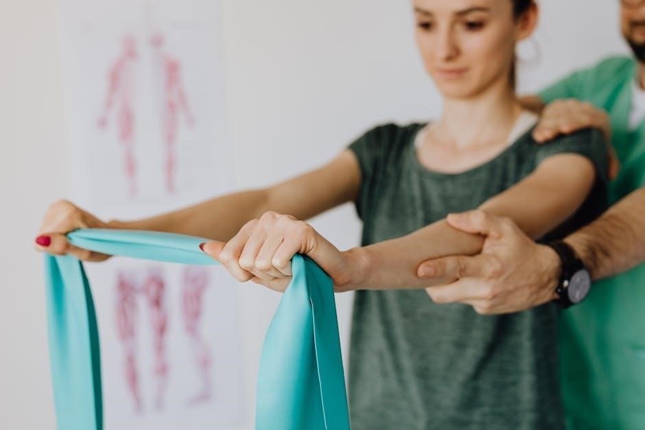
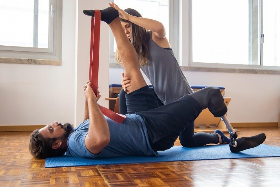
Targeted exercises are vital for correcting muscle imbalances contributing to ITBS. Clamshells strengthen the gluteus medius, crucial for hip stability and preventing excessive knee valgus. Perform 3 sets of 15-20 repetitions. Side-lying leg lifts further enhance gluteal strength; aim for 3 sets of 15-20 repetitions per leg.
Hip abduction exercises with a resistance band also target the gluteus medius. Single-leg squats improve strength and balance, challenging the hip and knee stabilizers. Start with assisted squats if needed. Step-downs from a low platform similarly build strength and control.
These exercises should be performed with proper form to avoid exacerbating the condition. Focus on controlled movements and avoid pain. Gradually increase resistance or repetitions as strength improves. A physical therapist can provide personalized exercise guidance and ensure correct technique, maximizing effectiveness and minimizing risk of re-injury. Consistency is key for long-term improvement.
Foam rolling the IT band, while often uncomfortable, can help release tension and improve flexibility. Start at the hip and slowly roll down to just above the knee, pausing on tender spots for 30 seconds. Repeat 2-3 times per leg. Self-massage using a lacrosse ball can target specific trigger points along the IT band and surrounding muscles.
Additionally, foam rolling the quadriceps, hamstrings, and glutes is crucial, as tightness in these areas can contribute to ITBS. Spend at least 30 seconds on each muscle group. Gentle, sustained pressure is more effective than aggressive rolling. Remember to breathe deeply during the process.
These techniques are best performed before and after exercise. While foam rolling doesn’t “lengthen” the IT band itself, it addresses the surrounding musculature. Combining foam rolling with stretching and strengthening exercises provides a comprehensive approach to ITBS management. Listen to your body and stop if you experience sharp pain.
Strengthening the muscles surrounding the hip and knee is vital for correcting biomechanical imbalances that contribute to ITBS; Gluteus medius strengthening is paramount; exercises like side-lying leg raises and clam shells (15-20 reps, 3 sets) help stabilize the pelvis.
Hip abductor exercises, including banded walks (20 steps each direction, 3 sets), further enhance pelvic control. Don’t neglect the quadriceps; squats and lunges (10-12 reps, 3 sets) build strength and endurance. Focus on maintaining proper form to avoid exacerbating the condition.
Hamstring curls (12-15 reps, 3 sets) and calf raises (15-20 reps, 3 sets) also contribute to overall lower limb stability. Progress gradually, increasing resistance as strength improves. Incorporate these exercises 2-3 times per week, allowing for adequate recovery between sessions. Consistent strengthening is key to long-term ITBS prevention.
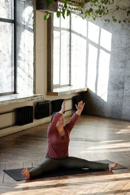
Stretching, while not directly lengthening the IT band itself, focuses on releasing tension in surrounding muscles. The iliopsoas stretch (hold 30 seconds, 3 reps per side) addresses hip flexor tightness, a common contributor to ITBS. A piriformis stretch (hold 30 seconds, 3 reps per side) can alleviate pressure on the sciatic nerve and improve hip mobility.
Quadriceps stretches, such as the standing quad stretch (hold 30 seconds, 3 reps per leg), are crucial for maintaining knee flexibility. Hamstring stretches, like the towel hamstring stretch (hold 30 seconds, 3 reps per leg), improve overall leg length and reduce strain.
Gentle calf stretches (hold 30 seconds, 3 reps per leg) also contribute to lower leg flexibility. Perform these stretches after warming up or after activity. Avoid bouncing or forcing the stretch; focus on a gentle, sustained hold. Consistency is key for improving flexibility and reducing ITBS symptoms.